It’s a question that crosses many patients’ minds when the dental assistant positions the lead apron and steps out of the room: “Is this really safe?” The concern is understandable. We’re conditioned to think of radiation as dangerous, and the precautions dental offices take—the heavy apron, the technician leaving the room—can feel unsettling even when everything is routine.
The good news? Dental x-rays are among the safest diagnostic tools in modern medicine, with radiation exposure so minimal it’s often compared to the background radiation you encounter during a short airplane flight or a few hours in the sun. But understanding why they’re safe—and why the benefits far outweigh the negligible risks—can transform dental x-rays from a source of anxiety into a reassuring part of your preventive care.
Understanding Radiation in Context
Radiation sounds scary because we associate it with nuclear disasters and cancer treatment. But radiation is actually everywhere. It’s a natural part of our environment, constantly surrounding us in forms we rarely think about.
Every day, you’re exposed to background radiation from cosmic rays, the soil beneath your feet, the building materials in your home, and even the foods you eat. Bananas contain potassium-40, a naturally radioactive isotope. Brazil nuts have radium. The granite countertops in many kitchens emit low levels of radiation continuously.
This constant exposure is measured in units called millisieverts (mSv). The average American receives about 3.1 mSv of background radiation annually just from living their normal life. To put dental x-rays in perspective:
- A single digital dental x-ray: approximately 0.005 mSv
- A full set of dental x-rays (typically 4 bitewings): approximately 0.02 mSv
- A panoramic dental x-ray: approximately 0.01-0.02 mSv
For comparison:
- Cross-country airplane flight: approximately 0.03 mSv
- Chest x-ray: approximately 0.1 mSv
- Mammogram: approximately 0.4 mSv
- CT scan of the head: approximately 2 mSv
- Annual background radiation: approximately 3.1 mSv
A full set of dental x-rays exposes you to less radiation than you’d receive during a flight from Los Angeles to New York. You’d need roughly 150 sets of dental x-rays to equal the radiation from a single head CT scan. The numbers put dental imaging firmly in the “negligible risk” category.
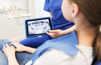
Digital X-Rays: A Quantum Leap in Safety
If traditional dental x-rays were already safe, digital x-rays—the technology used at modern practices like Torrance Dentistry—have made them even safer. Digital radiography reduces radiation exposure by up to 80-90% compared to conventional film x-rays.
The difference lies in the sensors. Traditional x-rays required enough radiation to expose photographic film. Digital sensors are far more sensitive, capturing detailed images with a fraction of the radiation dose. The images appear instantly on a computer screen, can be enhanced for better diagnosis, and are stored electronically rather than requiring chemical processing.
Beyond reduced radiation, digital x-rays offer several advantages:
- Immediate Results: There’s no waiting for film development. Your dentist can review images within seconds of taking them.
- Enhanced Imaging: Digital images can be magnified, adjusted for contrast, and manipulated to reveal details that might be missed on traditional film.
- Easy Sharing: If you need to see a specialist or transfer to another practice, your x-rays can be shared electronically rather than requiring physical copies.
- Environmental Benefits: No chemical processing means no hazardous waste from developing solutions.
Why Your Dentist Leaves the Room
This is perhaps the most misunderstood aspect of dental x-rays. If they’re so safe, why does the technician step behind a wall or leave the room entirely?
The answer isn’t that each individual x-ray is dangerous—it’s about cumulative exposure over time. Dental professionals may take dozens of x-rays every single day, year after year, for the duration of their careers. While a patient might receive a handful of x-rays annually, a dental technician could potentially be exposed to thousands.
The protective measures aren’t about the danger to you during your single appointment. They’re about protecting staff members from the additive effect of repeated low-level exposure over decades. The ALARA principle—As Low As Reasonably Achievable—guides radiation safety in healthcare, meaning we minimize exposure whenever possible, even when individual doses are already very low.
This is actually reassuring. The precautions exist not because dental x-rays are dangerous, but because the dental profession takes radiation safety so seriously that they protect against even theoretical long-term risks to their staff.
The Lead Apron Question
You’ve likely worn a heavy lead apron during dental x-rays your entire life. Interestingly, current guidelines from the American Dental Association and the FDA suggest that lead aprons are no longer considered necessary for most dental x-rays when modern equipment and techniques are used.
Modern dental x-ray machines produce highly focused, narrow beams that target only the specific area being imaged. Scatter radiation—the type that would affect other parts of your body—is minimal with digital equipment. Some studies suggest the lead apron provides more psychological comfort than actual radiation protection with contemporary technology.
That said, many dental practices continue using lead aprons because patients expect them and find them reassuring. There’s no harm in wearing one, and if it makes you more comfortable during your x-rays, that’s reason enough to use it. Thyroid collars remain recommended for certain types of x-rays, particularly for children and during panoramic imaging.
When and Why Dental X-Rays Are Necessary
Understanding why x-rays matter helps put any remaining concerns in proper perspective. The radiation exposure from dental x-rays is minimal; the consequences of undetected dental problems can be severe.
Dental x-rays reveal what visual examination cannot:
- Decay Between Teeth: Cavities that form where teeth touch are invisible to the naked eye until they’ve become quite large. X-rays catch them when they’re small and easily treated.
- Decay Under Existing Fillings: Old fillings can develop new decay underneath them, hidden from view but visible on x-rays.
- Bone Loss: Periodontal disease causes bone loss around tooth roots. By the time bone loss becomes clinically obvious, significant damage has occurred. X-rays detect early bone changes.
- Abscesses and Infections: Infections at the root tips of teeth may cause no symptoms initially but appear clearly on x-rays, allowing treatment before they spread or become painful emergencies.
- Impacted Teeth: Wisdom teeth and other teeth that haven’t erupted properly can cause problems for adjacent teeth. X-rays reveal their position and trajectory.
- Cysts and Tumors: Though rare, abnormal growths in the jaw can be detected on routine x-rays, sometimes before any symptoms appear.
- Root Problems: Fractures, resorption, and other root issues that would be impossible to diagnose otherwise.
The question isn’t whether dental x-rays carry risk—everything in medicine involves some level of risk-benefit analysis. The question is whether the benefit of early detection and prevention outweighs the minimal radiation exposure. For dental x-rays, the answer is clearly yes.
How Often Should You Have Dental X-Rays?
There’s no universal schedule that applies to everyone. X-ray frequency depends on your individual oral health status, risk factors, and dental history. A patient with no history of cavities, excellent home care, and healthy gums may need x-rays less frequently than someone with a history of decay or periodontal disease.
General guidelines suggest:
Bitewing X-Rays (for detecting decay between teeth):
- High-risk patients: every 6-12 months
- Moderate-risk patients: every 12-18 months
- Low-risk patients: every 24-36 months
Full Mouth Series or Panoramic X-Rays:
- New patients: typically at the initial comprehensive exam
- Established patients: every 3-5 years, or as needed based on clinical findings
Periapical X-Rays (for examining specific teeth):
- As needed to evaluate symptoms, monitor treatment, or investigate specific concerns
Your dentist determines appropriate x-ray frequency based on your unique situation. If you’re unsure why particular x-rays are recommended, ask. Understanding the reasoning helps you make informed decisions about your care.
Special Considerations: Children and Pregnancy
Parents often express extra concern about x-rays for their children, and pregnant women frequently wonder whether dental x-rays should be avoided entirely.
For Children: Pediatric dental x-rays use even lower radiation doses than adult x-rays, and children actually benefit significantly from dental imaging. Their mouths are changing rapidly, and x-rays help monitor tooth development, detect decay early (children are often at higher risk), and identify orthodontic issues before they become more complex.
The American Academy of Pediatric Dentistry supports appropriate use of dental radiography in children, emphasizing that the diagnostic benefits outweigh the minimal radiation exposure when x-rays are clinically indicated.
During Pregnancy: The American College of Obstetricians and Gynecologists and the American Dental Association both affirm that dental x-rays are safe during pregnancy. The radiation dose is extremely low, the beam is directed at the mouth rather than the abdomen, and lead aprons with thyroid collars provide additional protection.
That said, many dentists prefer to postpone routine x-rays until after pregnancy when possible, simply out of abundance of caution. Emergency or diagnostic x-rays shouldn’t be delayed due to pregnancy, however—untreated dental infections pose greater risks to both mother and baby than the negligible radiation from dental imaging.
What Torrance Dentistry Does to Maximize Safety
At Torrance Dentistry, patient safety guides every aspect of care, including diagnostic imaging. Dr. Steve Yabuno and Dr. Daniel Yabuno have invested in state-of-the-art digital radiography that minimizes radiation exposure while maximizing diagnostic quality.
The practice’s approach to x-ray safety includes:
- Modern Digital Equipment: Digital sensors require significantly less radiation than traditional film, reducing exposure by up to 80-90%.
- Individualized X-Ray Schedules: Rather than taking x-rays on a rigid schedule, the doctors at Torrance Dentistry determine imaging frequency based on each patient’s specific needs and risk factors.
- Proper Technique: Equipment is regularly calibrated and maintained, and staff follows established protocols to ensure optimal imaging with minimal exposure.
- Lead Aprons and Thyroid Collars: Protective equipment is available for patients who want additional reassurance.
- Strict Protocols: The practice adheres to all ADA and state guidelines regarding radiation safety.
With over 40 years of experience serving the South Bay community, Dr. Steve Yabuno has witnessed the evolution of dental imaging from traditional film to today’s digital technology. His commitment to staying current with advances in dental technology—including completing continuing education in the latest techniques—ensures that patients at Torrance Dentistry benefit from the safest, most effective diagnostic tools available.
The Bottom Line on Dental X-Ray Safety
Dental x-rays are safe. The radiation exposure is minimal—comparable to the background radiation you receive simply by existing in the world. Digital technology has made an already-safe procedure even safer. And the diagnostic benefits of dental imaging far outweigh the negligible risks.
Avoiding dental x-rays doesn’t make you safer—it makes you more vulnerable to undetected decay, infection, bone loss, and other problems that are far more consequential than the tiny amount of radiation involved in imaging. The real risk isn’t in the x-ray; it’s in what goes undiagnosed without one.
Schedule Your Visit at Torrance Dentistry
Dr. Steve Yabuno and Dr. Daniel Yabuno, along with their experienced team of specialists and hygienists, provide comprehensive dental care for families throughout Torrance, Redondo Beach, Lomita, and the surrounding South Bay communities. With on-staff periodontists, advanced technology including digital x-rays and intraoral cameras, and a commitment to patient education, Torrance Dentistry offers everything you need for optimal oral health in one convenient location.
The practice is located at 3500 Lomita Blvd, Suite 103, in Torrance. Hours are Monday through Thursday from 8:00 AM to 5:30 PM, and Friday through Saturday from 8:00 AM to 2:00 PM. Call (310) 530-7011 to schedule your appointment, or visit the website to learn more about services and request an appointment online.
Your questions about dental x-rays—or any aspect of your care—are always welcome. The team at Torrance Dentistry believes informed patients make the best decisions about their oral health, and they’re happy to discuss any concerns you may have.
Posted on behalf of
3500 Lomita Blvd #103
Torrance, CA 90505
Phone: (310) 530-7011
Email: info@torrancedentistry.com